Our recent paper, “Super-Resolution in Clinically Available Spinal Cord MRIs Enables Automated Atrophy Analysis”, published in the American Journal of Neuroradiology, explores an exciting advancement in how we analyze spinal cord atrophy in MS. Atrophy on magnetic resonance imaging (MRI) of the nervous system is thought to reflect the heath of nerve cells. The spinal cord is the part of the central nervous system that helps move limbs, carry sensations to the brain, control bladder and bowel function, and more. Spinal cord atrophy is thus closely tied to motor and sensory problems for people with MS. However, measuring spinal cord atrophy is hard. The types of scans required to make atrophy measurements are very high resolution, higher resolution than those collected in standard clinical practice, and there is tremendous time required from experts to analyze the images.
In our work, we introduce a new use of a tool called SMORE that uses artificial intelligence to enhance the resolution of the more typical low-resolution spinal cord MRIs acquired in clinical practice. This technique is particularly useful as it works without additional data or external training. Using SMORE, we can measure atrophy reliably, even when starting with low-resolution images.
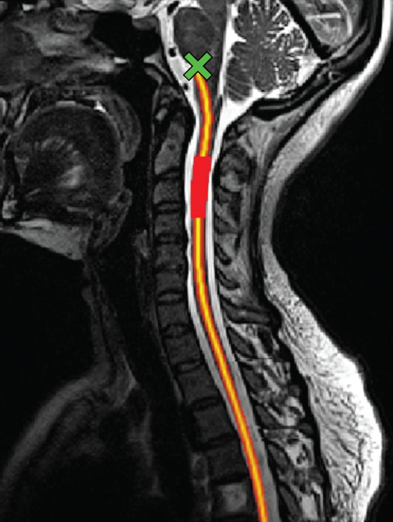
When comparing scans generated by SMORE to research-quality high-resolution scans, we found that they match well, with strong correlations in image quality and atrophy measurements. More importantly, after enhancing the quality of scans through SMORE, atrophy measures correlated better with clinical outcomes—such as walking speed and overall disease severity—than the original low-resolution scans. We also validated our approach using real-world datasets, confirming that SMORE-enabled measurements can accurately track spinal cord atrophy over time. This opens the door to large-scale, cost-effective studies on spinal cord atrophy using existing clinical imaging datasets, reducing the need for specialized research scans.
This work represents a significant step forward for MS research and care. We can bring advanced imaging analytics to a much broader audience by leveraging routinely available MRI scans and enhancing their resolution. For patients, this means more precise tracking of disease progression and potentially more personalized treatment plans. For researchers, it enables larger, more inclusive studies to understand the disease better and test new therapies. This study demonstrates how cutting-edge technology like SMORE can bridge the gap between clinical practice and research innovation, ultimately improving the lives of people living with MS.